


Canadian Medical Association Journalwww.cmaj.ca
- CMAJ December 6, 2005 vol. 173 no. 12 1441-1442
CMAJ December 6, 2005 vol. 173 no. 12
- © 2011 Canadian Medical Association or its licensors
- All editorial matter in CMAJ represents the opinions of the authors and not necessarily those of the Canadian Medical Association.
- Practice
Transient monocular visual loss and retinal migraine
+ Author Affiliations
The Case: A 40-year-old man was referred because of multiple events of transient monocular visual loss since adolescence. He described the events as small, translucent, grey-coloured spots similar to those seen after looking at a bright light. These visual defects affected both of his eyes equally in frequency but never simultaneously (i.e., each episode was monocular). Episodes lasted about 5–10 minutes, occurred 2–3 times a day on average and were often associated with migrainous headaches, which began within 30 minutes after the onset of the visual symptoms. The headaches were described as unilateral, pounding, lasting up to 4 hours, and sometimes associated with nausea, vomiting, photophobia and phonophobia. On 1 or 2 occasions the headaches also coincided with unilateral jaw and arm numbness, and scintillating scotoma. Although the headaches responded well to ibuprofen, the visual symptoms did not.
On examination, the patient's visual acuity was 20/20 (6/6) in both eyes, and the intraocular eye pressure was 14 mm Hg bilaterally (normal pressure 10–20 mm Hg). Gross examination of the visual fields by confrontation yielded normal findings, as did the rest of the ophthalmologic and the neurologic examinations. Comprehensive blood work revealed only mild hyperlipidemia. Electrocardiography demonstrated sinus rhythm, and neither carotid duplex Doppler ultrasonography nor transthoracic echocardiography revealed any abnormalities. Retinal migraine was diagnosed, and daily therapy with ASA and verapamil was started. The frequency of events of transient visual loss decreased from 2–3 attacks per day to 2–3 attacks per week.
Retinal migraines are transient monocular visual disturbances (scintillations, scotomas or blindness) that can occur simultaneously with migraine headaches or in a patient with a prior history of migraines. They occur because of hypoperfusion of either the eye or the optic nerve. This is in contrast to typical migraine with aura (previously known as classic migraine), which involves the cerebral cortex and is therefore associated with binocular visual phenomena. The International Headache Society's definition of retinal migraine is given in Box 1.1 The society's definition is limited because it does not account for patients who have visual symptoms without headaches or who have permanent visual scotomas; although rare, both of these presentations have been well documented in the literature.
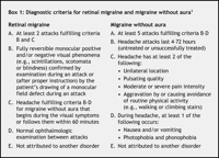
Box 1.
Retinal migraine affects about 1 of every 200 patients who have migraines. At the time of diagnosis, most patients are less than 40 years old. Nearly 30% have a past history of nonretinal migraine with or without aura, and 25% have a relative with retinal migraines.
Clinically, retinal migraine has a highly variable presentation. Some patients describe primarily negative symptoms (visual loss) consisting of black, grey, white or shaded areas of varying size that may appear instantaneously or gradually progress inward from the peripheral visual field. Others describe positive symptoms such as flashing lights or scintillating scotomas. Symptoms are always monocular. Most events are transient, lasting from 5–20 minutes, and may occur several times a day. When headaches occur in association with the visual changes, they may occur either during or after the visual disturbances.
Box 2.
Specific precipitants for attacks are often unclear. Although the natural history of retinal migraine has not been well studied, the prognosis appears to be similar to that of typical migraine with aura. Symptoms may go into permanent remission after several months or years, they may go into remission but recur at a future date, or they may persist lifelong.
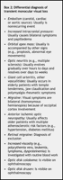
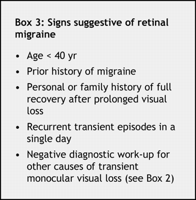
The diagnosis of retinal migraine is by exclusion. The differential diagnosis of transient monocular visual loss is given in Box 2. A careful history and physical examination are required, often in conjunction with appropriate investigations, including a complete blood count, erythrocyte sedimentation rate, prothrombin and partial thromboplastin times, electrocardiography, carotid duplex Doppler ultrasonography, transthoracic echocardiography and, if an arrhythmia is suspected, a Holter monitor. A work-up for hypercoagulable states is often indicated if the personal or family history is suggestive. Signs that should raise clinical suspicion of retinal migraine are listed in Box 3.
Box 3.
Management is often done in collaboration with a neurologist or an ophthalmologist. Formal baseline visual field testing is advisable; abnormalities may indicate a need for additional investigation or closer follow-up. If the episodes of transient visual loss have been infrequent, management is often conservative, with reassurance and simple follow-up. Although no guidelines are available, we recommend prophylactic therapy if the episodes are disabling or occur more than twice per week. Low-dose daily ASA therapy is well tolerated and has been reported anecdotally to be effective. Although randomized trials of therapies are lacking, anecdotal reports have suggested that calcium-channel blockers such as verapamil and nifedipine,2 β-blockers3 and inhaled amyl nitrate therapy4 may be effective for prophylaxis.
No comments:
Post a Comment